42 labels of the human brain
101 Labeled Brain Images and a Consistent Human Cortical Labeling ... Labeling the macroscopic anatomy of the human brain is instrumental in educating biologists and clinicians, visualizing biomedical data, localizing brain data for identification and comparison, and perhaps most importantly, subdividing brain data for analysis. Diagram Of Brain with their Labelings and Detailed Explanation - BYJUS The parietal lobe is found at the upper back of our brain. This lobe functions by controlling all our complex behaviours, including senses of vision, the sense of touch, spatial orientation and body awareness. It manages body position, movements, the perception of stimuli, orientation, handwriting and visuospatial processing. The Occipital Lobe
2,821 Labeled brain anatomy Images, Stock Photos & Vectors - Shutterstock Labeled brain anatomy royalty-free images 2,810 labeled brain anatomy stock photos, vectors, and illustrations are available royalty-free. See labeled brain anatomy stock video clips Image type Orientation Sort by Popular Healthcare and Medical Anatomy human brain brain organ medicine cerebellum cerebrum human body cerebral cortex Next of 29
Labels of the human brain
3D Brain This interactive brain model is powered by the Wellcome Trust and developed by Matt Wimsatt and Jack Simpson; reviewed by John Morrison, Patrick Hof, and Edward Lein. Structure descriptions were written by Levi Gadye and Alexis Wnuk and Jane Roskams . Solved Label the structures and lobes of the human brain by | Chegg.com Expert Answer. 97% (29 ratings) 1. Temporal lobe 2. Insula 3. Fronta …. View the full answer. Transcribed image text: Label the structures and lobes of the human brain by clicking and dragging the labels to the correct location. <--Anterior Posterior --> Precentral gyrus Temporal lobe Parieto-occipital sulcus Parietal lobe Lateral sulcus ... Brain: Anatomy, Pictures, Functions, and Conditions - Verywell Mind The cerebral cortex is the part of the brain that makes human beings unique. Functions that originate in the cerebral cortex include: Consciousness Higher-order thinking Imagination Information processing Language Memory Perception Reasoning Sensation Voluntary physical action 1 The cerebral cortex is what we see when we look at the brain.
Labels of the human brain. Frontiers | 101 Labeled Brain Images and a Consistent Human Cortical ... We introduce the Mindboggle-101 dataset, the largest and most complete set of free, publicly accessible, manually labeled human brain images. To manually label the macroscopic anatomy in magnetic resonance images of 101 healthy participants, we created a new cortical labeling protocol that relies on robust anatomical landmarks and minimal manual edits after initialization with automated labels ... Labeled Parts Of The Brain Illustrations, Royalty-Free Vector Graphics ... detailed anatomy of the human brain. detailed anatomy of the human brain. Illustration showing the medulla, pons, cerebellum, hypothalamus, thalamus, midbrain. Sagittal view of the brain. Isolated on a white background. Pineal gland anatomical cross section vector illustration... Brain Basics: Know Your Brain | National Institute of Neurological ... The brain can be divided into three basic units: the forebrain, the midbrain, and the hindbrain. The hindbrain includes the upper part of the spinal cord, the brain stem, and a wrinkled ball of tissue called the cerebellum ( 1 ). The hindbrain controls the body's vital functions such as respiration and heart rate. Labeled Brain Model Diagram | Science Trends The frontal lobe of the brain is responsible for our critical thinking, planning, reasoning, and problem-solving, as well as our experience of emotions. The rear portion of the frontal lobe is the motor cortex, which receives inputs from the other lobes and carries out the movements of the body associated with them.
The Human Brain Atlas - Michigan State University In this atlas you can view MRI sections through a living human brain as well as corresponding sections stained for cell bodies or for nerve fibers. The stained sections are from a different brain than the one which was scanned for the MRI images. Furthermore, for the stained sections, the brain was removed from the skull, dehydrated, embedded ... Labeled Diagrams of the Human Brain You'll Want to Copy Now The central core consists of the thalamus, pons, cerebellum, reticular formation and medulla. These five regions are the central areas that regulate breathing, pulse, arousal, balance, sleep and early stages of processing sensory information. The thalamus interprets the sensory information and helps determine what is good and bad. Human Brain Anatomy - Components of Human Brain with Images Human Brain Anatomy: The brain is composed of the complex network of billions of neurons that are arranged in a specific pattern which is vital to the essential functioning of this organ. Working on the principle of division of labour, different parts of brain are specialized for only specific tasks. The entire mass is enclosed inside a hard ... Brain Anatomy and How the Brain Works | Johns Hopkins Medicine Gray and white matter are two different regions of the central nervous system. In the brain, gray matter refers to the darker, outer portion, while white matter describes the lighter, inner section underneath. In the spinal cord, this order is reversed: The white matter is on the outside, and the gray matter sits within.
The human brain: Parts, function, diagram, and more - Medical News Today The brain is the human body's control system, and is part of the central nervous system (CNS). It connects to the spine and controls personality, movement, breathing, and other crucial processes ... Brain: Atlas of human anatomy with MRI - e-Anatomy - IMAIOS Choroid plexus of fourth ventricle Choroid plexus of lateral ventricle Choroid plexus of third ventricle Choroidal fissure Cingulate gyrus Cingulate sulcus Cingulum Circular sulcus of insula Cistern of lamina terminalis Cistern of lateral cerebral fossa Claustrum Collateral eminence Collateral sulcus Parts of the brain: Learn with diagrams and quizzes | Kenhub Labeled brain diagram. First up, have a look at the labeled brain structures on the image below. Try to memorize the name and location of each structure, then proceed to test yourself with the blank brain diagram provided below. Labeled diagram showing the main parts of the brain. The Human Brain | Brain and Cognitive Sciences | MIT OpenCourseWare Image of a cortex with colored labels of the regions resposible for various perceptual and cognitive functions. (Courtesy of the instructor.) Course Description This course surveys the core perceptual and cognitive abilities of the human mind and asks how they are implemented in the brain.
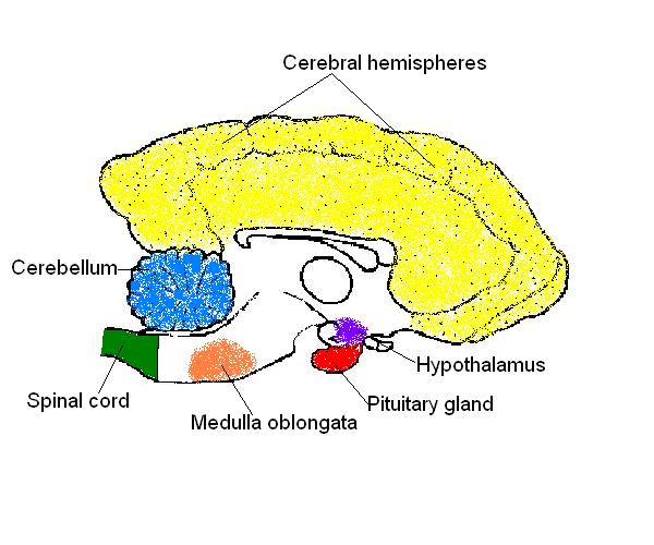
The Anatomy and Physiology of Animals/Nervous System Worksheet/Worksheet Answers - WikiEducator
Amazon.com: XINDAM 3D Human Brain with Labels Anatomical Model ... This item: XINDAM 3D Human Brain with Labels Anatomical Model Paperweight (Laser Etched) in Crystal Glass Ball Science Gift (Included LED Base) $66.99 Brain 11 Ounce Ceramic Coffee Mug (WC462M) $16.98 Anatomic Brain Specimen Coasters (Set of 10) - Neuroscience Gifts, Gifts for Medical Student Gifts Brain Decor Human Anatomy Gifts

my red crayon: put your thinking caps on.
Amazon.com: brain model labeled 1-16 of 487 results for "brain model labeled" RESULTS Price and other details may vary based on product size and color. Amazon's Choice Learning Resources Cross-section Brain Model - 2 Pieces, Ages 7+ Brain Anatomy Model, Brain Functions Model, Human Anatomy for Kids, Foam Brain Model 1,619 $1999 Get it as soon as Fri, Sep 2
What are some good online resources (ideally providing good visuals and animation to aid in the ...
The Human Brain - Visible Body The brain gives us self-awareness and the ability to speak and move in the world. Its four major regions make this possible: The cerebrum, with its cerebral cortex, gives us conscious control of our actions. The diencephalon mediates sensations, manages emotions, and commands whole internal systems. The cerebellum adjusts body movements, speech ...
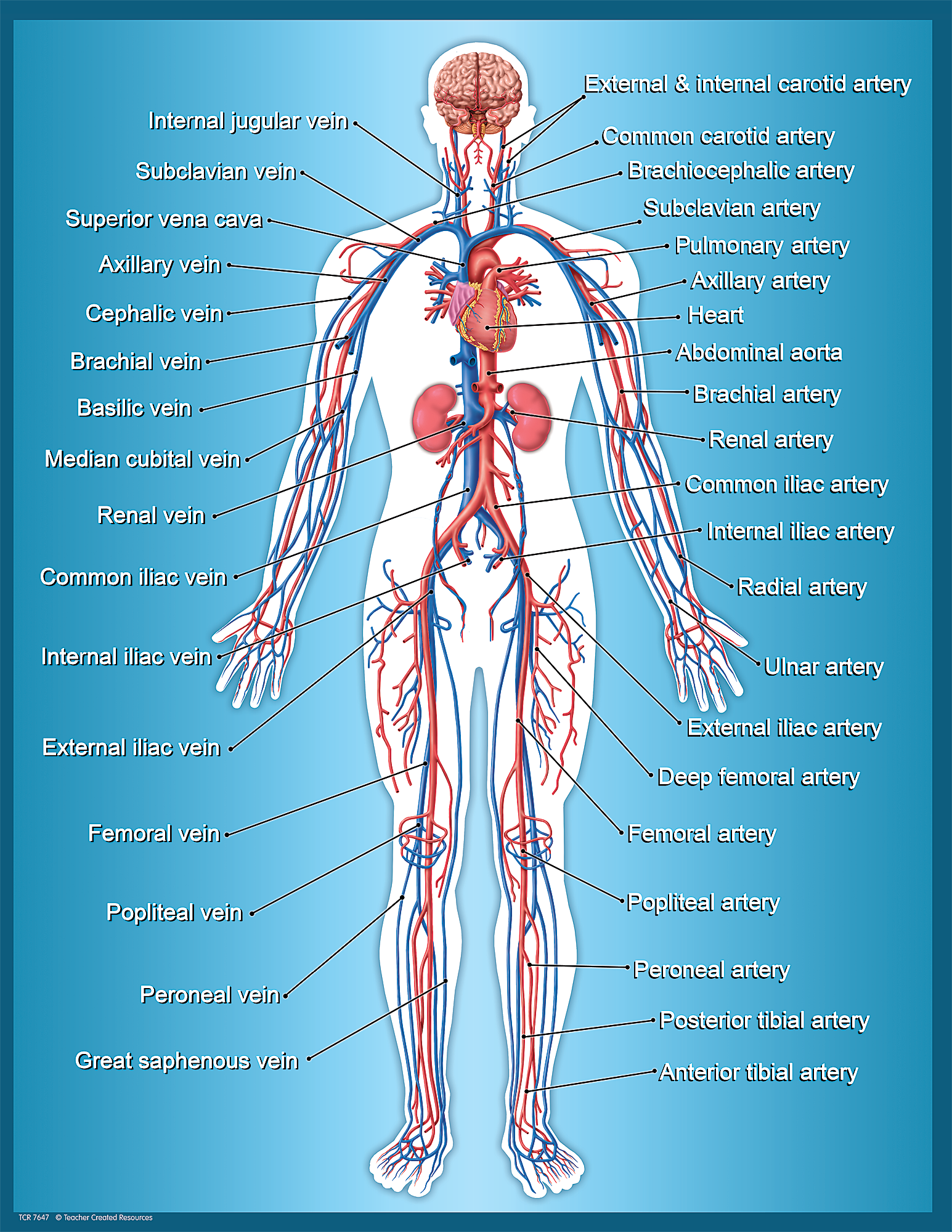
Circulatory System Chart - TCR7647 | Teacher Created Resources
Human Brain - Structure, Diagram, Parts Of Human Brain - BYJUS The cerebrum is the largest part of the brain. It consists of the cerebral cortex and other subcortical structures. It is composed of two cerebral hemispheres that are joined together by heavy, dense bands of fibre called the corpus callosum. The cerebrum is further divided into four sections or lobes:

neuropathology blog: February 2013
Nervous System - Label the Brain - TheInspiredInstructor.com Nervous System - Label the Brain Nervous System - Brain Name: Choose the correct names for the parts of the brain. ( 1) (2) (3) (4) (5) (6) (7) (8) ( 9) This brain part controls thinking. (10) This brain part controls balance, movement, and coordination. (11) This brain part controls involuntary actions such as breathing, heartbeats, and digestion.

33 Brain Anatomy Quiz Label - Labels Database 2020
Human brain - Wikipedia The brainstem includes the midbrain, the pons, and the medulla oblongata. Behind the brainstem is the cerebellum ( Latin: little brain ). [8] The cerebrum, brainstem, cerebellum, and spinal cord are covered by three membranes called meninges. The membranes are the tough dura mater; the middle arachnoid mater and the more delicate inner pia mater.

Pin on Neuroscience-brain education
Brain (Human Anatomy): Picture, Function, Parts, Conditions ... - WebMD • The cortex is the outermost layer of brain cells. Thinking and voluntary movements begin in the cortex. • The brain stem is between the spinal cord and the rest of the brain. Basic functions like...

Human A&P lab unit 2 flashcards | Quizlet
Brain - Human Brain Diagrams and Detailed Information - Innerbody The brainstem is made of three regions: the medulla oblongata, the pons, and the midbrain. A net-like structure of mixed gray and white matter known as the reticular formation is found in all three regions of the brainstem. The reticular formation controls muscle tone in the body and acts as the switch between consciousness and sleep in the brain.

Midsagittal section of brain Quiz
Labeled Human Brain Illustrations, Royalty-Free Vector ... - iStock Browse 109 labeled human brain stock illustrations and vector graphics available royalty-free, or start a new search to explore more great stock images and vector art. Brain functions vector illustration. Labeled explanation organ... Brain functions vector illustration. Labeled explanation head organ parts scheme.

27 Label The Brain Anatomy Diagram - Wiring Database 2020
Anatomy of the Brain: Structures and Their Function - ThoughtCo The brain and spinal cord are the two main structures of the central nervous system. There are three major divisions of the brain. They are the forebrain, the midbrain, and the hindbrain. Key Takeaways The forebrain, the midbrain, and the hindbrain are the three main parts of the brain.
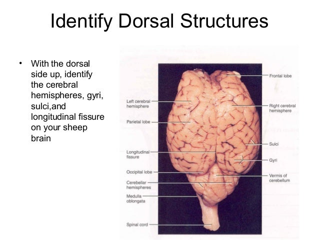
Sheep+brain+dissection
brain labeling blank anatomy brain quiz labeling cortex cerebral label parts game areas labels ch13 fig01 mc creative. Label The Brain Answers - Juleteagyd juleteagyd.blogspot.com. paintingvalley. Label The Brain Worksheets (SB11585) - SparkleBox | Human Brain Diagram . brain label diagram drawing worksheet labels worksheets diagrams labelled ...

Anatomy of the Brain anatomy poster | A well, Charts and My love
Brain: Anatomy, Pictures, Functions, and Conditions - Verywell Mind The cerebral cortex is the part of the brain that makes human beings unique. Functions that originate in the cerebral cortex include: Consciousness Higher-order thinking Imagination Information processing Language Memory Perception Reasoning Sensation Voluntary physical action 1 The cerebral cortex is what we see when we look at the brain.
30 Human Brain With Label - Labels For You
Solved Label the structures and lobes of the human brain by | Chegg.com Expert Answer. 97% (29 ratings) 1. Temporal lobe 2. Insula 3. Fronta …. View the full answer. Transcribed image text: Label the structures and lobes of the human brain by clicking and dragging the labels to the correct location. <--Anterior Posterior --> Precentral gyrus Temporal lobe Parieto-occipital sulcus Parietal lobe Lateral sulcus ...

Anatomy - teaching resource | Lt + LabStation | ADInstruments
3D Brain This interactive brain model is powered by the Wellcome Trust and developed by Matt Wimsatt and Jack Simpson; reviewed by John Morrison, Patrick Hof, and Edward Lein. Structure descriptions were written by Levi Gadye and Alexis Wnuk and Jane Roskams .

Amazon.com: The Brain Anatomy Poster: Science Lab Anatomy Classroom Supplies… | Brain anatomy ...

Image Gallery | MBF Bioscience

Bald Worm's Blog: Year 5 - Planning: The Fab Four Golden Rules
Post a Comment for "42 labels of the human brain"