44 human eye diagram without labels
diagram of eye with labels Eye and Ear Models. 11 Pictures about Eye and Ear Models : Picture Of the Eye Labeled Elegant Human Eye Anatomy for Kids | Human, Eye Diagram Without Labels | via Anatomy Pictures Gallery if… | Flickr and also Picture Of the Eye Labeled Elegant Human Eye Anatomy for Kids | Human. Eye And Ear Models . ear anatomy eye ... Activity Sheet 1: How the Eyes Work | Human eye diagram, Teaching ... Description Use these simple eye diagrams to help students learn about the human eye. Three differentiated worksheets are included: 1. Write the words using a word bank 2. Cut and paste the words 3. Write the words without a word bank Labels include: eyebrow, eyelid, eyelashes, pupil, iris, and sclera.
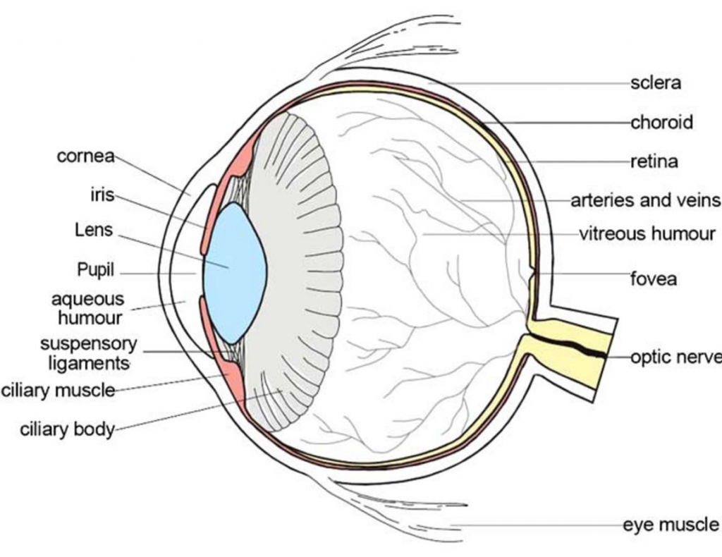
PDF Eye Anatomy Handout - National Eye Institute of light entering the eye. Lens: The lens is a clear part of the eye behind the iris that helps to focus light, or an image, on the retina. Macula: The macula is the small, sensitive area of the retina that gives central vision. It is located in the center of the retina. Optic nerve: The optic nerve is the largest sensory nerve of the eye.

Human eye diagram without labels
Heart Diagram Labeled Igcse : The Human Eye Edexcel Igcse Biology ... This diagram shows the parts of the eye. 9 the diagram shows the apparatus used to demonstrate the action of amylase on starch. Veins carry deoxygenated blood and wastes from the tissues to the liver and heart. The heart's muscle is constantly active. 60,758 Human eye anatomy Images, Stock Photos & Vectors - Shutterstock Find Human eye anatomy stock images in HD and millions of other royalty-free stock photos, illustrations and vectors in the Shutterstock collection. Thousands of new, high-quality pictures added every day. BYJUS BYJUS
Human eye diagram without labels. anatomy human eye Anatomy eye labeled human body sclera physiology models spinal google optic nerve notes ik 4d nerves bing. ... eye diagram unlabeled unlabelled anatomy human 11e3 medical. Extreme Close-Ups Of The Human Eye - Neatorama ... Egg Emerging From An Ovary - Weird Picture Archive . hand human inside egg tendons skin without ... The Human Eye | Boundless Physics | | Course Hero Diagram of the Human Eye: The cornea and lens of an eye act together to form a real image on the light-sensing retina, which has its densest concentration of receptors in the fovea and a blind spot over the optic nerve.The power of the lens of an eye is adjustable to provide an image on the retina for varying object distances. Layers of tissues with varying indices of refraction in the lens ... Human Ear Diagram - Bodytomy The Structure of Human Ear. Helix: It is the prominent outer rim of the external ear. Antihelix: It is the cartilage curve that is situated parallel to the helix. Crus of the Helix: It is the landmark of the outer ear, situated right above the pointy protrusion known as the tragus. Auditory Ossicles: The three small bones in the middle ear ... PDF Parts of the Eye - National Eye Institute | National Eye Institute Eye Diagram Handout Author: National Eye Health Education Program of the National Eye Institute, National Institutes of Health Subject: Handout illustrating parts of the eye Keywords: parts of the eye, eye diagram, vitreous gel, iris, cornea, pupil, lens, optic nerve, macula, retina Created Date: 12/16/2011 12:39:09 PM
Anatomy of the eye: Quizzes and diagrams | Kenhub Take a look at the diagram of the eyeball above. Here you can see all of the main structures in this area. Spend some time reviewing the name and location of each one, then try to label the eye yourself - without peeking! - using the eye diagram (blank) below. Unlabeled diagram of the eye human face diagram and label human face diagram and label The Skull - Scientific Publishing we have 8 Pics about The Skull - Scientific Publishing like Facial Anatomy & Proportions - Hair & Makeup Artist Handbook, picture front of the eye without labels clipart - Clipground and also Head Outline Clip Art at Clker.com - vector clip art online, royalty. Here it is: The Eyes (Human Anatomy): Diagram, Optic Nerve, Iris, Cornea ... - WebMD The front part (what you see in the mirror) includes: Iris: the colored part. Cornea: a clear dome over the iris. Pupil: the black circular opening in the iris that lets light in. Sclera: the ... Human eye - Wikipedia The human eye is a sensory organ, part of the sensory nervous system, that reacts to visible light and allows us to use visual information for various purposes including seeing things, keeping our balance, and maintaining circadian rhythm.. The eye can be considered as a living optical device.It is approximately spherical in shape, with its outer layers, such as the outermost, white part of ...
Eye anatomy: A closer look at the parts of the eye In a number of ways, the human eye works much like a digital camera: Light is focused primarily by the cornea — the clear front surface of the eye, which acts like a camera lens. The iris of the eye functions like the diaphragm of a camera, controlling the amount of light reaching the back of the eye by automatically adjusting the size of the ... Label Parts of the Human Eye - University of Dayton Label Parts of the Human Eye. Select One Anterior Chamber Ciliary Body Cornea Fibrous Tunic Iris Lateral Rectus Muscle Lens Medial Rectus Muscle Optic Disk Optic Nerve Pupil Retina Vascular Tunic Vitreous Nerve. Select One Anterior Chamber Ciliary Body Cornea Fibrous Tunic Iris Lateral Rectus Muscle Lens Medial Rectus Muscle Optic Disk Optic ... human eye diagram with labels Eye With Labels Clip Art at Clker.com - vector clip art online, royalty. 10 Images about Eye With Labels Clip Art at Clker.com - vector clip art online, royalty : Muscles of the Human Eyeball | ClipArt ETC, 35 Label The Structure Of The Eye - Labels Database 2020 and also 35 Label The Structure Of The Eye - Labels Database 2020. Eye Anatomy: Parts of the Human Eye - Vision Center The following are parts of the human eyes and their functions: 1. Conjunctiva. The conjunctiva is the membrane covering the sclera (white portion of your eye). The conjunctiva also covers the interior of your eyelids. Conjunctivitis, often known as pink eye, occurs when this thin membrane becomes inflamed or swollen.
Human Anatomy Review and Question Database
File:Schematic diagram of the human eye no.svg - Wikimedia Original upload log []. This image is a derivative work of the following images: File:Schematic diagram of the human eye en.svg licensed with PD-self 2008-02-02T01:33:45Z Jakov 508x516 (54267 Bytes) suspensory ligament, arrow was wrong; 2008-01-31T16:48:11Z Jakov 508x516 (54263 Bytes) xml-Cleanup; 2007-01-25T03:10:10Z Rhcastilhos 508x516 (42056 Bytes) {{Information |Description=Schematic ...
parts of the eyes clipart 20 free Cliparts | Download images on Clipground 2021
eyeball diagram labeled Eye Diagram Without Labels | Via Anatomy Pictures Gallery If… | Flickr . fluke. Model Of Human Eye - YouTube . eye human. Eye section cross anatomy eyes. Ear worksheet diagram inner anatomy animals physiology senses answers parts eye special labelled worksheets ossicles auditory outer pinna middle canal.

10 Best Images of Label Ear Diagram Worksheet - Blank Rock Cycle Diagram, Frog Dissection ...
Eye Diagram Teaching Resources | Teachers Pay Teachers The Human Eye Overview Reading Comprehension and Diagram Worksheet. by. Teaching to the Middle. 63. $1.50. Zip. This passage briefly describes the human eye (900-1000 Lexile). 14 questions (matching and multiple choice) assess students' understanding. Students label a diagram of 6 parts of the eye. I've included a color and BW version, as well ...

Sinus Diagram. Human Anatomy (Without Labels Version) Stock Photo 171111779 : Shutterstock
Eye Anatomy: Parts of the Eye and How We See Behind the anterior chamber is the eye's iris (the colored part of the eye) and the dark hole in the middle called the pupil. Muscles in the iris dilate (widen) or constrict (narrow) the pupil to control the amount of light reaching the back of the eye. Directly behind the pupil sits the lens. The lens focuses light toward the back of the eye.

Blank Ear Diagram | Human ear diagram, Ear anatomy, Ear diagram
Eye Diagram - Differentiated Worksheets and EASEL Activities - Pinterest Description Use these simple eye diagrams to help students learn about the human eye. Three differentiated worksheets are included: 1. Write the words using a word bank 2. Cut and paste the words 3. Write the words without a word bank Labels include: eyebrow, eyelid, eyelashes, pupil, iris, and sclera. UPDATE: I've updated this product to ...

picture front of the eye without labels clipart - Clipground
2,781 Labeled brain anatomy Images, Stock Photos & Vectors - Shutterstock Find Labeled brain anatomy stock images in HD and millions of other royalty-free stock photos, illustrations and vectors in the Shutterstock collection. Thousands of new, high-quality pictures added every day.

Tolle The Diagram Labeled Ideen Menschliche Anatomie - Blank Brain Diagram Lobes | Transparent ...
ear diagram without labels eye parts human diagram without labels clipart front anatomy eyes simple body science label ear students activities primary printable worksheets Drag each label to the appropriate location on this diagram of the. Ear human drawing draw parts easy way diagram science getdrawings paintingvalley. Pin on kids science experiments/ crafts/school ideas
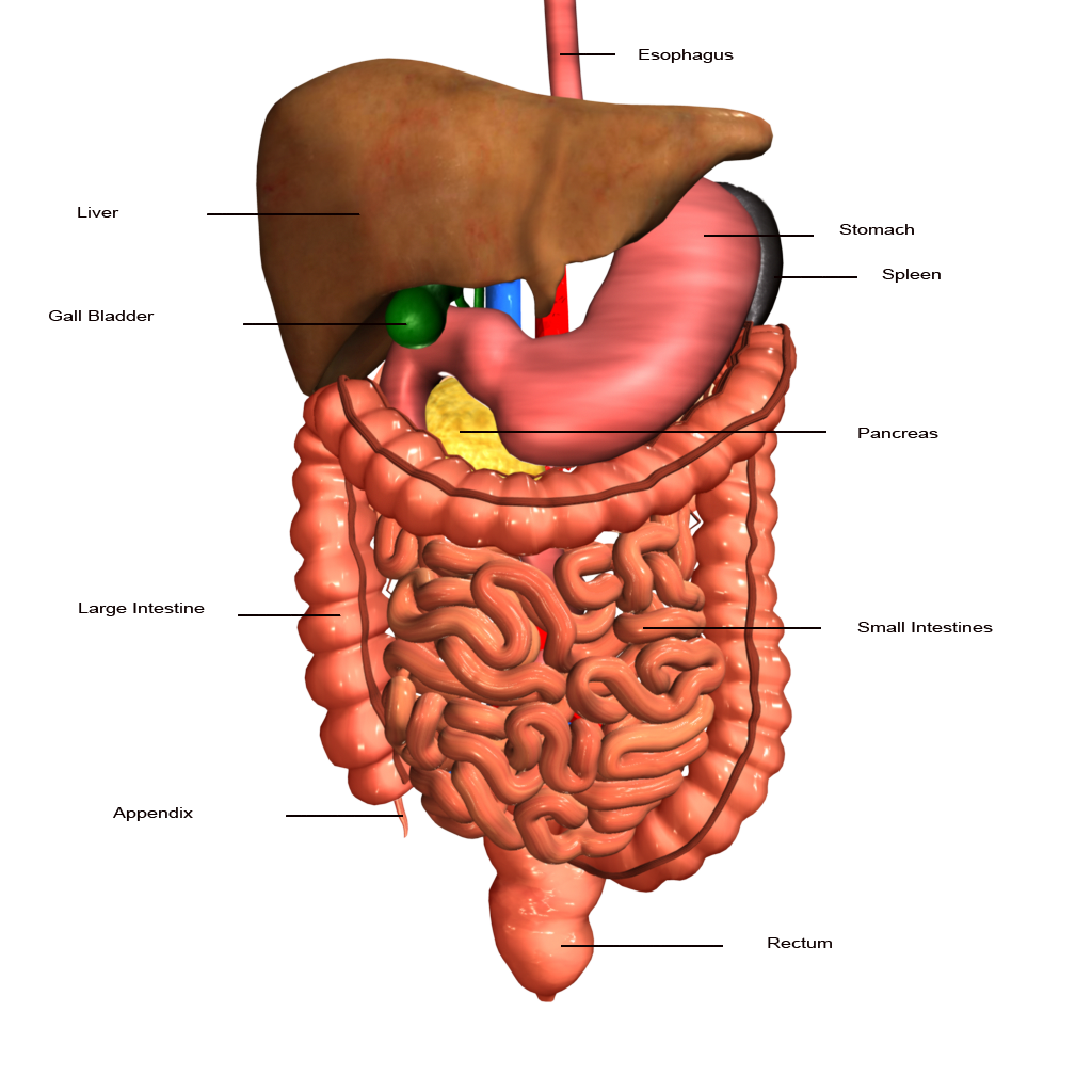
Transparent Digestive System Without Labels ~ news word
Eye Diagram With Labels and detailed description - BYJUS A brief description of the eye along with a well-labelled diagram is given below for reference. Well-Labelled Diagram of Eye The anterior chamber of the eye is the space between the cornea and the iris and is filled with a lubricating fluid, aqueous humour. The vascular layer of the eye, known as the choroid contains the connective tissue.
Post a Comment for "44 human eye diagram without labels"