45 picture of the eye with labels
Solved B с A E F D Match the following parts of the eye with - Chegg Science. Anatomy and Physiology. Anatomy and Physiology questions and answers. B с A E F D Match the following parts of the eye with the labels in the picture above. A Iris F Cornea В. Ciliary Muscles G Optic Nerve C Lens E Retina Aqueous and Vitreous Fluid. Question: B с A E F D Match the following parts of the eye with the labels in the ... Eye With Labels Clip Art at Clker.com - Free Clip Art & Images Download Clker's Eye With Labels clip art and related images now. Multiple sizes and related images are all free on Clker.com.
Human Eye Diagram - Human Body Pictures & Images - Science for Kids Photo description: This human eye diagram gives an excellent overview of the human eye. The cross section features labeled parts such as the iris, pupil, cornea, lens, retina, choroid, optic disc, optic nerve and fovea. For more information on eyes, check out our range of interesting human eye facts.

Picture of the eye with labels
› helpful-garden-redirectHelpful Garden Redirect — Foresight Montessori Oh No! We know this isn’t what you were looking for… …And we are sorry for this inconvenience. All of the links from the original Helpful Garden blog and google ... Eye Anatomy Diagram - EnchantedLearning.com Definitions : Aqueous humor - the clear, watery fluid inside the eye. It provides nutrients to the eye. Astigmatism - a condition in which the lens is warped, causing images not to focus properly on the retina. Binocular vision - the coordinated use of two eyes which gives the ability to see the world in three dimensions - 3D. Labelling the eye — Science Learning Hub In this interactive, you can label parts of the human eye. Use your mouse or finger to hover over a box to highlight the part to be named. Drag and drop the text labels onto the boxes next to the eye diagram If you want to redo an answer, click on the box and the answer will go back to the top so you can move it to another box.
Picture of the eye with labels. Packaging | Custom Boxes Wholesale | Create Your Own ... Design marvelous custom packaging and custom printed boxes with Emenac Packaging to create a unique brand identity and glorify your product presentation. Get premium quality custom boxes wholesale at extremely affordable prices that fit your budget with no minimum quantity restrictions, fast turnaround time and free shipping anywhere is USA Label Functions of Parts of the Human Eye Functions of the Parts of the Eye. Select the correct label for the function of each part of the eye. The image is taken from above the left eye. Click on the Score button to see how you did. Incorrect answers will be marked in red. Eye Diagram With Labels and detailed description - BYJUS A brief description of the eye along with a well-labelled diagram is given below for reference. Well-Labelled Diagram of Eye The anterior chamber of the eye is the space between the cornea and the iris and is filled with a lubricating fluid, aqueous humour. The vascular layer of the eye, known as the choroid contains the connective tissue. Label Eye Printout - EnchantedLearning.com Label the Eye Diagram. Human Anatomy. Read the definitions, then label the eye anatomy diagram below. Cornea - the clear, dome-shaped tissue covering the front of the eye. Iris - the colored part of the eye - it controls the amount of light that enters the eye by changing the size of the pupil. Lens - a crystalline structure located just behind ...
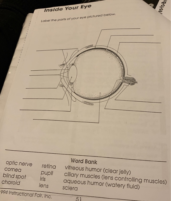
Eye Anatomy: 16 Parts of the Eye & Their Functions - Vision Center The lens of the eye (or crystalline lens) is the transparent lentil-shaped structure inside your eye. This is the natural lens. It is located behind the iris and to the front of the vitreous humor (vitreous body). The vitreous humor is a clear, colorless, gelatinous mass that fills the gap between the lens and the retina in the eye. Pin on Ylli - Pinterest Use this simple eye diagram for primary students as they learn about the human eye. Two differentiated worksheets included: one with a word bank and one without. Words to label: eyebrow, eyelid, eyelashes, pupil, iris, and sclera. Find this Pin and more on Ylli by Kirsi Liuska. Eye Anatomy Diagram. Ear Diagram. Science Student. What is an eye mark and why do I need it? - Consolidated Label An 'eye mark' (also known as 'eye spot') is a small rectangular printed area located near the edge of the printed flexible packaging material. A sensor on the form-fill-seal (FFS) machine reads the eye mark to identify packaging material, control the material's position, and coordinate the separation and cutting of the flexible packaging material. en.wikipedia.org › wiki › Motion_Picture_AssociationMotion Picture Association - Wikipedia The Motion Picture Association represents the interests of the six international producers and distributors of filmed entertainment. To do so, they promote and protect the intellectual property rights of these companies and conduct public awareness programs to highlight to movie fans around the world the importance of content protection.
Human eye diagram Images, Stock Photos & Vectors - Shutterstock Human eye diagram royalty-free images 6,729 human eye diagram stock photos, vectors, and illustrations are available royalty-free. See human eye diagram stock video clips Image type Orientation Sort by Popular Biology Healthcare and Medical human eye anatomy eye retina medicine visual perception cone cell pupil Next of 68 The Eye - Science Quiz - GeoGuessr The Eye - Science Quiz: Our eyes are highly specialized organs that take in the light reflected off our surroundings and transform it into electrical impulses to send to the brain. The anatomy of the eye is fascinating, and this quiz game will help you memorize the 12 parts of the eye with ease. Light enters our eyes through the pupil, then passes through a lens and the fluid-filled vitreous ... 20 Different Ways to Draw the Eye - Improve Drawing Pen and water is a spontaneous technique that will add the element of chance into your drawings. First, establish the underlying form of the eye with a pen. Draw simple shapes to illustrate where the main outline of the eye. Using a water-soluble pen, draw with a sketching line to map out the main features. PDF Parts of the Eye - National Eye Institute | National Eye Institute Eye Diagram Handout Author: National Eye Health Education Program of the National Eye Institute, National Institutes of Health Subject: Handout illustrating parts of the eye Keywords: parts of the eye, eye diagram, vitreous gel, iris, cornea, pupil, lens, optic nerve, macula, retina Created Date: 12/16/2011 12:39:09 PM
Street Art By ArtFlyMovie: NYCHOS THE WEIRD - Some Extracts of his Amazing Work
Human eye diagram, Eye anatomy, Diagram of the eye - Pinterest The medulla is made of number of pyramidal structures containing renal tubules or Nephrons projecting into the cavity towards the inner region of kidney called pelvis.This is the region where renal artery and renal vein enter the kidney. Free end of pelvis shows cup like depressions called calyces. Renal pyramids of medulla project into these ...

Eclectitude: Bleigiessen Glass Sculpture - Thomas Heatherwick, Artist
en.wikipedia.org › wiki › Sparkle:_Original_MotionSparkle: Original Motion Picture Soundtrack - Wikipedia Sparkle: Original Motion Picture Soundtrack is the soundtrack album for the 2012 Sony/TriStar Pictures film Sparkle, a remake of the 1976 film of the same name. The album was released through Sony Music Entertainment 's RCA Records on July 31, 2012.

Eye Label
Transverse Section Of Eye Anatomy With Labels High-Res Vector Graphic ... Transverse section of eye anatomy with labels. - stock illustration. Transverse section of eye anatomy with labels. Buy the print. Get this image in a variety of framing options at Photos.com.
Vector label for free download about (6,449) vector label. sort by newest first page (95/95)
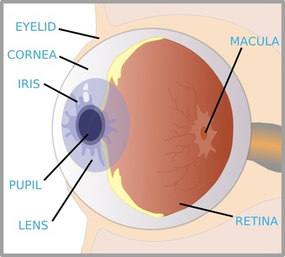
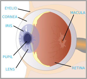
Eye Anatomy Detail Picture Image on MedicineNet.com Picture of Eye Anatomy Detail The eye is our organ of sight. The eye has a number of components which include but are not limited to the cornea, iris, pupil, lens, retina, macula, optic nerve, choroid and vitreous. Cornea: clear front window of the eye that transmits and focuses light into the eye.

Label Eye Diagram
Label Parts of the Human Eye - University of Dayton Parts of the Eye Select the correct label for each part of the eye. The image is taken from above the left eye. Click on the Score button to see how you did. Incorrect answers will be marked in red.
+(Left+Eye)+(2).jpg)
Aquarious' TLC Fan Blog: TLC's 'Waterfalls' Video Photo Shots
justagirlandherblog.com › make-labels-in-microsoftHow to Make Pretty Labels in Microsoft Word - Abby Lawson 5. “Group” your label. The last thing I like to do when I get the label the way I want it is to group it together. This allows me to move it around the page as one unit rather than trying to move each piece individually.
33 Label The Parts Of The Eye - Labels For You
Printable Eye Images | Etsy Check out our printable eye images selection for the very best in unique or custom, handmade pieces from our prints shops.

Missing Beats of Life: Eyes HD Wallpapers and Images
Structure and Functions of Human Eye with labelled Diagram The External Structure of an Eye. Sclera: It is a white visible portion. It is made up of dense connective tissue and protects the inner parts. Conjunctiva: It lines the sclera and is made up of stratified squamous epithelium. It keeps our eyes moist and clear and provides lubrication by secreting mucus and tears.
Aquarious' TLC Fan Blog: TLC's 'Waterfalls' Video Photo Shots
Illustration Picture of Anatomical Structures - Eye These parts include the cornea, iris, pupil, lens, retina, retinal blood vessels, and the vitreous body. Cornea: The cornea makes up the front-center part of the eye's outer wall. The cornea bends light, focusing it on the retina. People who wear contact lenses place their lenses on the cornea. Iris: The iris is the colored part of the eye.

Melanie Hicks Hot Pics and Bio | Picture Perfect
› best-sticker-printers12 Best Sticker Printer For Labels, Stickers, And Photos In 2022 May 04, 2022 · 10 sheets of 2×3 inch paper- for fast pictures or stickers, you may use the adhesive back picture paper with the peel-and-stick backing that comes with the kit. Features: Customize photos with borders and stickers, print from social media or your camera roll, view your photos in augmented reality, easily view photo libraries from the app ...
Eye With Labels clip art (111109) Free SVG Download / 4 Vector
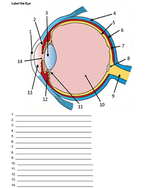
Label the Eye - The Biology Corner Label the Eye. Shannan Muskopf December 30, 2019. This worksheet shows an image of the eye with structures numbered. Students practice labeling the eye or teachers can print this to use as an assessment. There are two versions on the google doc and pdf file, one where the word bank is included and another with no word bank for differentiation.
30 Label Eye - Labels For Your Ideas
The Human Eye (Eyeball) Diagram, Parts and Pictures The eyeball is a round gelatinous organ that contains the actual optical apparatus. It is approximately 25 mm in diameter and sits snugly in the orbit where six muscles control its movement. The eyeball has three layers, each of which has several important structures that are essential for the sense of vision. Wall of the Eyeball

World's Beautiful things around us !: Beautiful nature| Eye cooling pictures every body loves to ...
PDF Eye Anatomy Handout - National Eye Institute of light entering the eye. Lens: The lens is a clear part of the eye behind the iris that helps to focus light, or an image, on the retina. Macula: The macula is the small, sensitive area of the retina that gives central vision. It is located in the center of the retina. Optic nerve: The optic nerve is the largest sensory nerve of the eye.

Label an Eye
Eye anatomy: A closer look at the parts of the eye The eye's crystalline lens is located directly behind the pupil and further focuses light. Through a process called accommodation, this lens helps the eye automatically focus on near and approaching objects, like an autofocus camera lens. ... The retina acts like an electronic image sensor of a digital camera, converting optical images into ...

Sports Day / Fun Run Poster | Free Early Years & Primary Teaching Resources (EYFS & KS1)
A Picture of the Eye - WebMD Your eye is a slightly asymmetrical globe, about an inch in diameter. The front part (what you see in the mirror) includes: Iris: the colored part. Cornea: a clear dome over the iris. Pupil: the ...
Post a Comment for "45 picture of the eye with labels"